Infectious Disease Case Study Part II
The Brooklyn International High School
Summer Research Program for Science Teachers
Summer 2006
Course: Living Environment (New York State Regents Curriculum)/Biology
Grade Level: 9th, 10th or 11th grades
Unit: Genetics, Biotechnology
Objectives: Students will be able to…
· List the steps an epidemiologist takes to detect a pathogen.
· Explain the steps in the polymerase chain reaction (PCR).
· Analyze the results from a gel electrophoresis.
· Describe how the polymerase chain reaction (PCR) and gel electrophoresis can be used to detect pathogens.
Introduction: With the growing fear of bioterrorism and a pandemic flu, scientists are developing new diagnostic tools to allow for rapid and sensitive detection of pathogens. This lab is based on a real outbreak of E.coli 0157:H7 that occurred in Japan in 1996. Before implementing this case study, students should have some background in biotechnology including an understanding of gel electrophoresis and PCR. It would also be helpful, though not essential, for students to have some background in pathogens and disease transmission.
Materials:
glue or tape (1 per pair)
scissors (1 per pair)
copies of handout (1 per student)
Time Required: 2 one-hour periods
Procedure:
New York State Math, Science, and Technology Learning Standards:
*Standard 1: Analysis, Inquiry and Design – Students will use mathematical analysis, scientific inquiry, and engineering design, as appropriate, to pose questions, seek answers, and develop solutions
*Standard 4: Science – Students will understand and apply scientific concepts, principles, and theories pertaining to the physical setting and living environment and recognize the historical development of ideas in science
*Standard: Interdisciplinary Problem Solving – Students will apply the knowledge and thinking skills of mathematics, science, and technology to address real-life problems and make informed decisions
National Science Learning Standards:
*Content Standard A: As a result of activities in grades
9-12, all students should develop abilities necessary to do scientific inquiry
and understanding about scientific inquiry
*Content Standard C: As a result of activities in grades 9-12, all students should develop understanding of the molecular basis of heredity, and matter, energy, and organization in living systems
*Content Standard E: As a result of activities in grades
9-12, all students should develop abilities of technological design and
understandings about science and technology
*Content Standard F: As a result of activities in grades 9-12, all students should develop understanding of science and technology in local, national, and global challenges
*Content Standard G: As a result of activities in grades 9-12, all students should develop understanding of the nature of scientific knowledge
In 1996, several students in Japan became ill with the following symptoms: diarrhea, abdominal pain, and in some cases, intestinal bleeding and kidney failure. After several tests, they were diagnosed with a bacterial infection caused by eating food contaminated with E. coli O157:H7. E. coli O157:H7 is one of the most virulent (highly infectious) pathogens known to enter the world food supply.
The symptoms were not specific enough to convince scientists definitively of the cause of the infection. So the scientists had to use specific tools to come to an accurate diagnosis of the cause of the disease. In this case, the scientists had to work very quickly because the children were young and therefore more likely to die from dehydration.
Directions: Imagine that you are a researcher investigating the cause of this outbreak.
Work with your colleagues to put the following procedure in order. Write the number next to the appropriate step.
|
|
PCR PCR is performed so that many copies of the bacterial DNA are available to test to determine the type of strain.
|
|
|
Data Analysis E. coli O157:H7. |
|
|
Isolate Bacteria Cells Then the scientists isolate the bacteria from the
sample. There are many other substances present in the stool sample. For
example, a stool sample would contain fecal matter, water, cells (including
human DNA), and other waste products in addition to the infectious bacteria. |
|
|
Collect Samples The scientists have to take samples from the sick children. The samples could be from the stool or vomit of the patients.
|
|
|
Gel Electrophoresis The PCR product is loaded into a gel so the product can
be visualized. |
|
|
Extract DNA from Bacteria Cells The bacterial cells are lysed (opened) to release the DNA. Of course when the bacteria cell membrane is opened, many other molecules and organelles are released. The DNA is separated from these other molecules through centifugation.
|
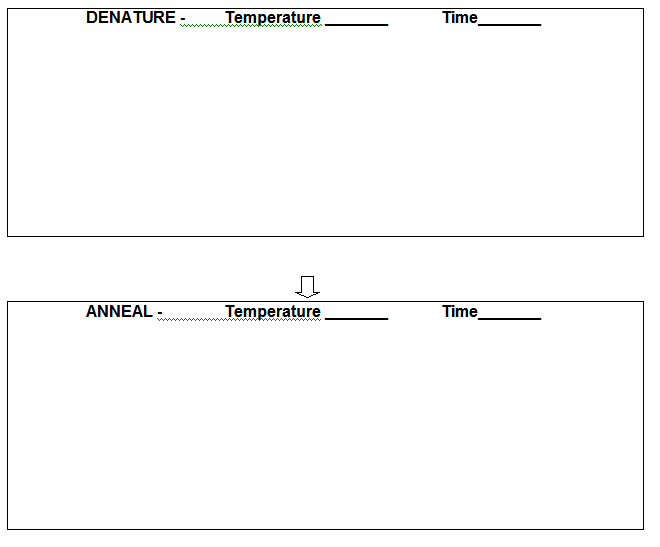
Directions:
1.
Cut out a double strand of E. coli
O157:H7
DNA.
2. Paste the double stranded DNA in the box labeled “Denature.”
Be sure to cut the strands
apart to show that the high temperature denatures the DNA.
3. Cut out a second double strand of E. coli O157:H7
DNA.
4. Paste the denatured double strand of DNA in the box labeled
“Anneal.”
5. Cut out the primers.
6. Paste the primers along the inside of each of the strands of DNA.
The underlined
sections represent areas where the primers will bind.
7. Cut out a third double strand of DNA and
place it in the box labeled “Extend.”
8. Cut out the primers and paste
them along side the DNA strands.
9. Continue to add on to the complementary sequence of the DNA strand by choosing
nucleotides from the dNTP
bottle. Cross out each nucleotide as you use it.
10. Analyze your pictures and write a caption under each one to explain what is happening.
11. Now that you have a large sample of DNA amplified, you can analyze it using gel electrophoresis to diagnose the disease. Lane A contains the DNA ladder, Lane B contains the patient’s sample, and Lane C contains a negative control (an uninfected individual).
The molecular weight of the PCR product (E. coli O157:H7 DNA with primers) should be 200-600bp. What is your diagnosis? Explain your answer.
Target DNA: E. coli O157:H7
Microtube of DNTPs:
Thermal Cycler - PCR Machine


PART I.
You are a researcher working at the Greene Infectious Disease Laboratory in New York City. A terrorist organization claims to have contaminated the subway system with the highly infectious bacteria, anthrax. The MTA immediately evacuated the subway, stopping all of the trains and requesting that the passengers take buses instead. You are part of an elite team of investigators whose task it is to determine whether this is a hoax or not.
PART II.
Now you are back in the lab with the samples that you collected from the subway. In the lab you have lots of equipment available to help you determine whether or not anthrax is present in the samples. You have a centrifuge, gel electrophoresis, PCR machine, primers, etc.
1.
What molecules (besides anthrax) would you expect to find in your subway
samples?
2.
With so many contaminants in your sample, how will you test specifically
for anthrax?
3.
If there is anthrax present in your sample, what would you use to
increase the concentration so that you have many copies available to test?
PART III.
You search the internet and order specific primers for anthrax PCR called PA and CAP primers. Through PCR you are able to produce many copies of this specific region of anthrax DNA, but you still have one more step to do before you can determine whether or not anthrax is present. You must run the PCR product on a gel for analysis. According to the primer information, if anthrax is present your PCR product will have two bands: one at 409 bp and another at 98 bp.
1.
How can your PCR product help you to identify the presence of anthrax?
2. Draw a picture of results that would indicate anthrax is present.
3. Draw a picture of your results that would indicate the absence of anthrax.